
AI Algorithm
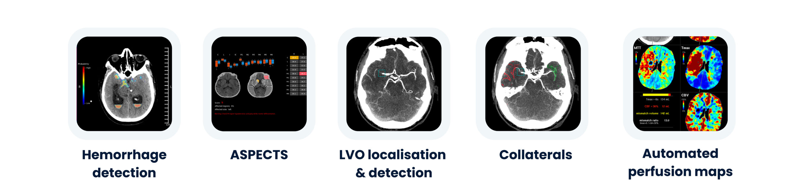
StrokeViewer’s AI Portfolio
Our advanced AI portfolio is purpose-built for stroke teams, seamlessly integrating cutting-edge technology into your workflow. From triage to treatment our AI-driven platform supports your stroke team every step of the way.
Automated Vessels Tracking
Our AI is built to accurately reconstruct the vasculature and trace precisely along the blood vessels until any potential thrombus is detected. This unique approach ensures fewer LVOs are missed
Suboptimal Scanning Quality
Trained on the renowned MR CLEAN and several other datasets that include patients across the world, so our AI can:
- Process thicker slices
- improve signal-to-noise-ratio;
- accept images from less advanced scanners
- Detect the phase of acquisition for quality assurance
High Sensitivity and Accuracy
Recent clinical studies show that our AI stroke algorithms’ high sensitivity and specificity give excellent accuracy, which is key to providing patients the treatment they deserve. Results are always visible, so any decisions are left to your own observation.
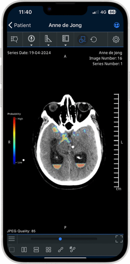
Detects subtle ICH & SAH hemorrhages
Hemorrhage detection
This AI stroke algorithm detects subtle bleeds and notifies your team immediately. Results are displayed via a binary contour mask and multicolored likelihood map for easy interpretation.
- Detection of subtle intracranial and subarachnoid hemorrhagic stroke
- Sensitivity of 92% (95%-confidence interval: 88%-95%) and a specificity of 100% (95%-confidence interval: 98%-100%) for cases ≥ 1mL*
- Auto-segmentation and volumetric measurements displayed through color-coded overlay.
* Instruction For Use (NIC-220193) based on internal validation data.
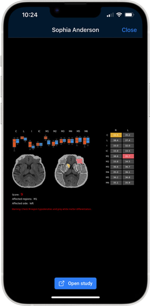
Automating ASPECTS
ASPECTS
Output of a standardized ASPECT score in minutes. With automatic detection of hypodensity on NCCT scans, you can quickly interpret results and make the best treatment decision.
- Alignment of the brain for easier reading of the scan
- Automatic identification of the ASPECTS regions on NCCT
- Delivers results with high consistency and reliability*
* Internal reproducibility report (NIC-230030); 2023
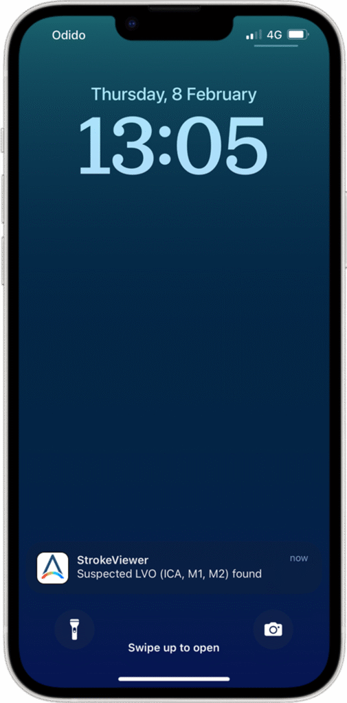
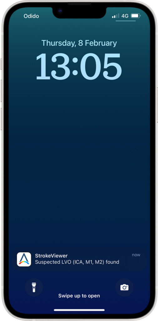
Reducing the number of undetected LVO’s
LVO detection
You and your team are instantly notified of a suspected Large Vessel Occlusion (LVO). The notification takes you straight to the patient timeline, where you can enter and store key clinical information.
- Access to results via your mobile device, email or PACS to diagnose an LVO patient faster
- Occlusion detection (including distal M2 branch)
- Performs with an overall sensitivity of 91%* and specificity of 86%* for ICA, M1 and M2 segments.
* IFU (NIC-220193)
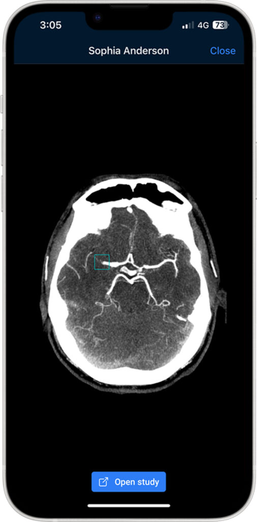
Precisely locating the LVO
LVO localisation
Your team is immediately notified of the precise location of a suspected occlusion. The notification takes you directly to the scan, helping you save time and reduce the risk of missing an LVO.
- Results are visible for your own interpretation
- LVO identifier to pinpoint LVO location
- You are navigated to the relevant slices containing the occlusion
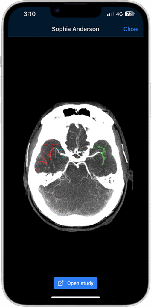
Quantitative measurements of the collateral circulation
Collaterals
Visualization of the vessels combined with thrombus location helps you determine whether the patient will benefit from endovascular therapy.
- Collateral status is the only proven treatment effect modifier currently in the clinical guidelines*
- You are informed of the acquisition phase for quality assurance
- The automated score is given as a quantitative collateral score and percentage of contralateral hemisphere, reducing interobserver variability
* A.M.M. Boers et al. AJNR 2018
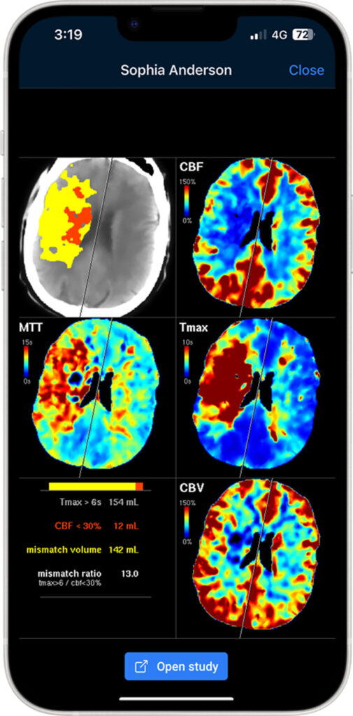
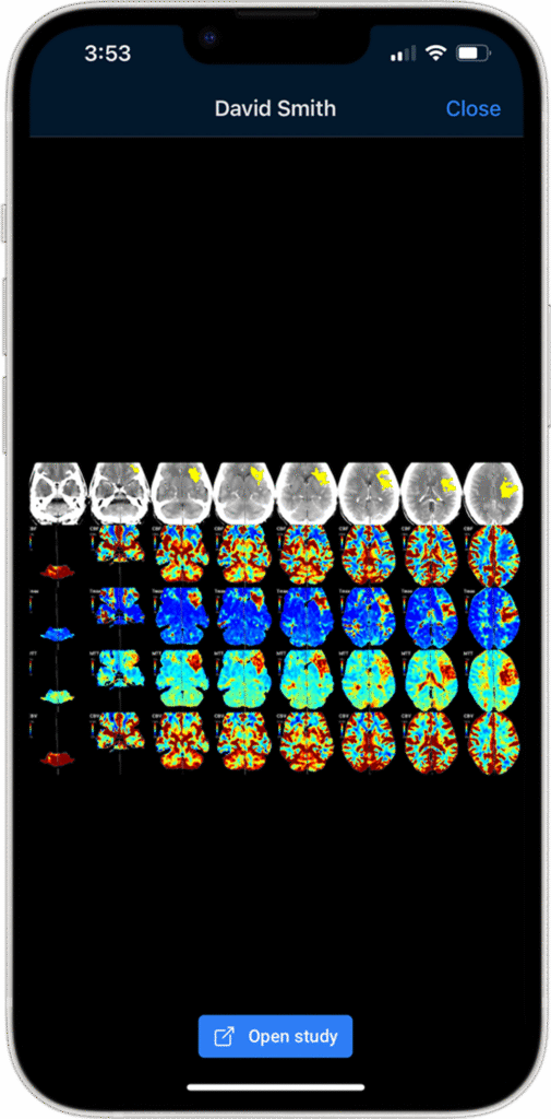
Automated quantification
Automated perfusion analysis
Quantifying reduced flow volume and mismatch volume help determine the amount of brain tissue that could be salvaged with endovascular treatment.
- Available for CT and MR
- Easy-to-interpret multicolored perfusion maps visible through our DICOM Viewer
- Automated AIF, VOF and motion correction to standardize results
Detects subtle ICH
Hemorrhage triaging
A real-time triage tool that helps healthcare provided quickly flag suspected acute intracranial Hemorrhage on head non-contrast CT scans.
- Detection of subtle subarachnoid and intraparenchymal, Epidural, Subdural and intraventricular hemorrhages.
- Sensitivity of 91.4%, Specificity of 97.5% and Accuracy of 95.6% for ICH detection*
- Case-level notifications are provided in case of suspected positive findings.
* Internal clinical validation study, 2023
Reducing the number of undetected LVO’s
LVO triaging
You and your team are instantly notified of a suspected Large Vessel Occlusion (LVO). The notification takes you directly to the patient scan, which you can help make informed decisions.
- Access to results via your mobile device, email or PACS.
- Sensitiviy of 91.3% and Specifity of 85.9% for occlusion detection.*
*Images were obtained primarily on GE scanners. Limited number of scans used from Siemens, Toshiba and Philips.
Automated quantification
CT Perfusion Analysis
- StrokeViewer offers multicolored perfusion maps through our user-friendly DICOM Viewer.*
- Perfusion volumes are calculated according to configurable Tmax and CBF thresholds
- Automated AIF and motion correction to standardize results
*Based on a survey conducted in 2021 among board-certified radiologists, neurologists and
other users