Extreme reward from a stroke workflow that impacts many patients
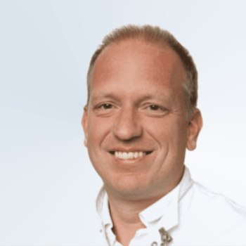
Dr. Wouter de Monyé is the Medical Director of Radiology at Spaarne Gasthuis. Having graduated from the University of Utrecht in 1996, de Monyé went on to carry out research at Leiden University Medical Center and trained as a radiologist at Leiden University Medical Center and Royal Liverpool University Hospital.
Why do you do what you do?
My motto, first and foremost, is to make people better. It’s a broad ambition, but importantly it’s not just patients but people I strive to better. That’s why I love to teach and train within the hospital. I want to help my fellow medical colleagues come to patient diagnoses faster and more efficiently.
What was your first job?
Funnily enough, cleaning a hospital when I was 15 years old. My mother was a nurse and set me up waxing those long corridors. As a child, I wanted to be a pilot, but this soon developed into the desire to become a flying doctor. You need to become a doctor first, and the aviation part comes later.
In the process, I discovered that radiology was my calling. I built computers on the side for a bit of extra money during my studies but soon had to give this up when I started my research at Leiden University.
What has been the highlight of your career so far?
Becoming the Medical Director of my department. This has allowed me to influence systems and processes, and improve the circumstances we work in as doctors. At some point, you think you’re doing your job and you’re a professional, but ultimately it’s the same process time and time again.
If you manage to get a helicopter view, you can see structural changes that could benefit not only one person but a cohort of patients. I find being able to influence such change extremely rewarding.
What is the most challenging aspect of your job?
It is getting the diagnosis right, particularly in acute situations. As a radiologist, you need to be able to ascertain what’s normal and abnormal and stick to it. But you also need to be able to know when to convey doubt. That’s the tricky part, especially when you don’t have time for a colleague to have a second look. Sometimes doctors put you on the spot – is it this or is it this? You find yourself having to give black or white answers in situations that are often pretty grey.
Do you have any coping mechanisms for dealing with the stress of your profession?
That’s difficult to answer. I guess realizing life can be over in the blink of an eyelid makes you focus on trying to build its most important elements, like good relationships and friendships.
Why is timely diagnosis so urgent in stroke?
The challenge to get things right is critical. Stroke is pretty common, and its impact can be huge – everlasting, for the rest of a patient’s life. Quick diagnosis is key, but more and more research suggests even in cases with a larger diagnosis time interval, there is still benefit from intra-arterial thrombolysis.
So having been proven in the MR CLEAN trial that in acute situations, this type of treatment option works, I think we’re now working towards expanding this approach to more patients. We’ll be seeing more situations whereby a possible clot has a chance to be treated in this novel way. I think the MR CLEAN trial gets more relevant every year.
Are there any challenges in the current stroke workflow?
Getting the knowledge to the image or the image to the knowledge. Doing the actual scans is no longer a problem. These days the CT scanners are able to produce high-quality images in a very short amount of time. The challenge is interpretation. Those many images need to be analyzed by an expert. Such professionals (ideally a neuroradiologist) are not always on call, and a patient’s scan will often have to be interpreted by colleagues with less professional experience.
Why is improving image sharing so vital?
A waste of time is a waste of brain. If we can get an image to an expert as quickly as possible, that can be life-altering for a patient. If a colleague needs help with interpretation, the current standard way of sharing images is difficult if you’re relying on your old-fashioned PACS system.
You receive a call when you’re out, and you have to go home, load up your computer, get the images up; this can easily lose you 15 minutes, which is absolutely critical. The shorter the door-to-needle time, the better the result.
With StrokeViewer, I can immediately receive a patient’s CT scan on my mobile phone wherever I am. That saves time by enabling me to interpret the high-quality images on the spot. It’s amazing, and it’s something I’ve never experienced with the regular PACS system. Seamless image sharing makes everything so much easier.
What are your thoughts on the added value of assisting image interpretation via artificial intelligence?
Assisting interpretation of CT images via artificial intelligence brings the bottom of quality up. It raises the bar. If, for whatever reason (timing, location, etc.), a less experienced radiologist is interpreting the scans – which is common – AI can help them perform like an experienced radiologist. That’s something that is really becoming clear from StrokeViewer and is so helpful.
How would you describe the current relationship between radiology and artificial intelligence? And how do you see this progressing?
In radiology, we’ve had digital images for quite a long time, more than 15 or 20 years. Even in the early stages, there were various computer-aided detection promises. But it’s only very recently, in the past 3 or 4 years, that deep learning and artificial intelligence make possible what was promised all that time ago.
I’ve worked with systems that have tried to do similar things but in a poor way. As with any relationship, it takes time to build a relationship of complete trust. And similarly, with any new technology, AI will need to prove itself in clinical practice. But we are currently seeing real-life applications of artificial intelligence, and it is amazing how fast my colleagues are beginning to rely on and trust it. It’s definitely going in the right direction.
Nicolab improves clinical practice with innovative and trusted AI.
Discover our StrokeViewer, an AI-powered clinical decision support system, offering a complete assessment of relevant imaging biomarkers within 3 minutes.

